Conulariida
Conulariida is a poorly understood fossil group that has possible affinity with the Cnidaria (jellyfish, gorgonian, coral, sea anemone). Members of the Conulariida are commonly referred to as conulariids and appear in the fossil record from the Ediacaran in the late Proterozoic (635-541ma) (Ivantsov and Fedonkin, 2002) (Van Iten et al., 2004) and extends without significant break through numerous major mass extinctions. The Conulariids finally disappear from the fossil record during the Late Triassic Period (~245 million years ago). In North America, conulariids are generally more common in rocks of Ordovician and Carboniferous age.
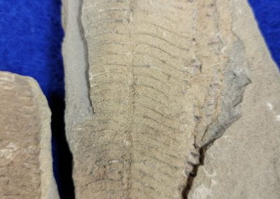
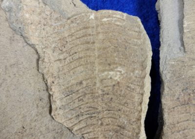
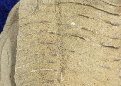
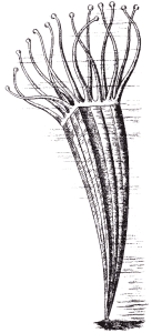
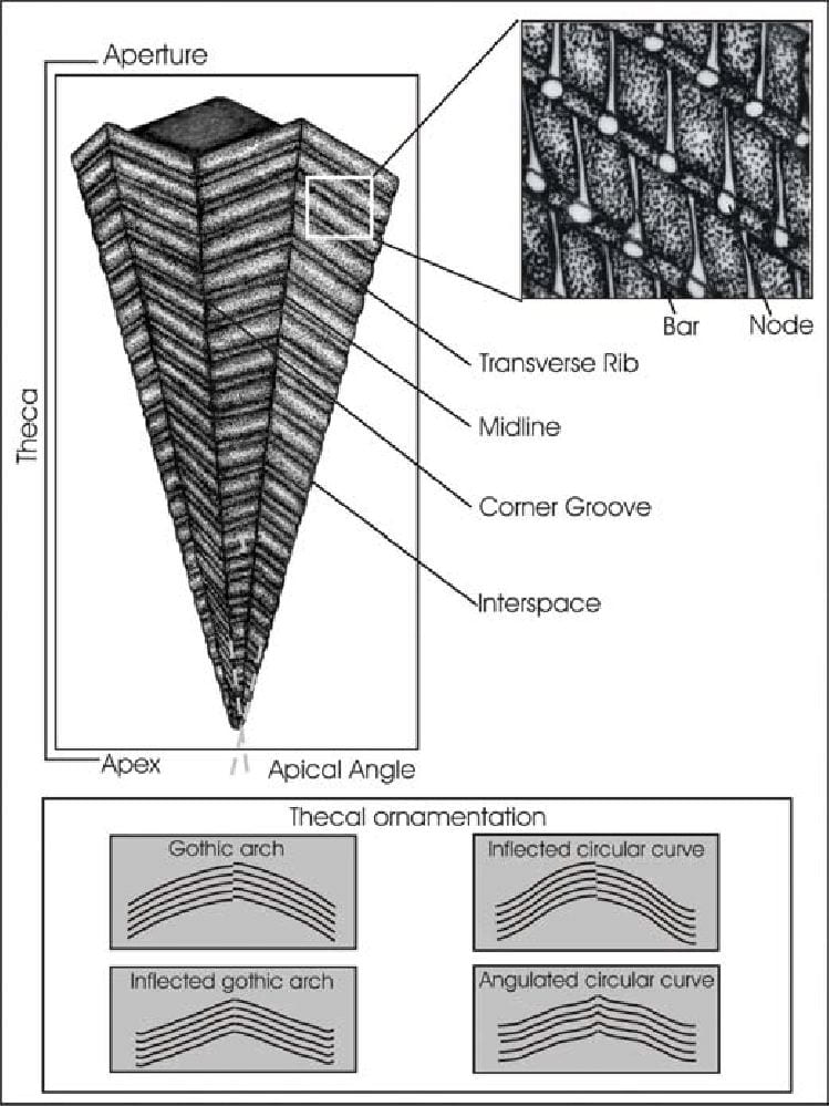
The conulariids are fossils preserved as shell-like structures made up of rows of calcium phosphate rods (transverse ribs), resembling an ice-cream cone with fourfold symmetry, usually four prominently-grooved corners (sulcus). However, there are some examples exhibiting three or six faces. New rods (transverse ribs) were added as the organism grew in length; the rib-based growth falsely gives the fossils a segmented appearance. Exceptional soft-part preservation has revealed that soft tentacles protruded from the wider end of the cone (aperture), and a holdfast from the pointed end (apex) attached the organisms to hard substrate. The prevailing reconstruction of the organism has it look superficially like a sea anemone sitting inside an angular, hard cone held perpendicular to the substrate. Conulariid shell is composed of a carbonate-rich apatite. The fine structure of their shell comprises multiple micro-, meso-, and macro-lamellae of alternately organic-rich and organic-poor layers (Ford, 2009). Observations of the organic matrix revealed that fibrous structures bridge the gaps left by the dissolution of the organic-poor microlamellae, connecting the organic-rich microlamellae. That description is analogous to Chapman and Werner’s (1972) description of a coronate scyphozoan periderm microstructure. This observation is another line of evidence strengthening the case for a conulariid and coronate scyphozoan relationship (Ford, 2009). Conulariids are thought to be most closely related to the Scyphozoa, or the “true jellyfish”. The current accepted taxonomy is that conulariids were modified descendants of a thecate cnidarian ancestor.
Conulariids apparently lived only in marine waters, such as the oceans and inland seas. Fossils are commonly found in rocks representing offshore, even anoxic, marine benthic environments. This has led some scientists to infer that these animals may have drifted planktonically for some or all of their lives, ultimately being buried in the anoxic sediments beneath the oxic waters in which they lived. However, basic functional considerations (such as the great weight of the shell) make such interpretations difficult to maintain.
Conulariids produced pearls within their shells, similar to the way molluscs such as oysters, other pelecypods, and some gastropods do today. These pearls give a clue as to the internal anatomy of the conulariid animal. But due to their calcium phosphate composition, their crystal structure, and their extreme age, these pearls tend to be rather unattractive for use in or as decorative objects.
What can we say about conulariids? They had elongated, pyramidal exoskeletons, made up of rows of calcium phosphate ribs. Most were square or rectangular in cross section, with prominent grooves at the corners. They lived attached to hard objects by a flexible stalk, and often lived in groups. Presumably they were filter feeders; how they reproduced is not known.
Location of Collection of Three Conulariid Samples
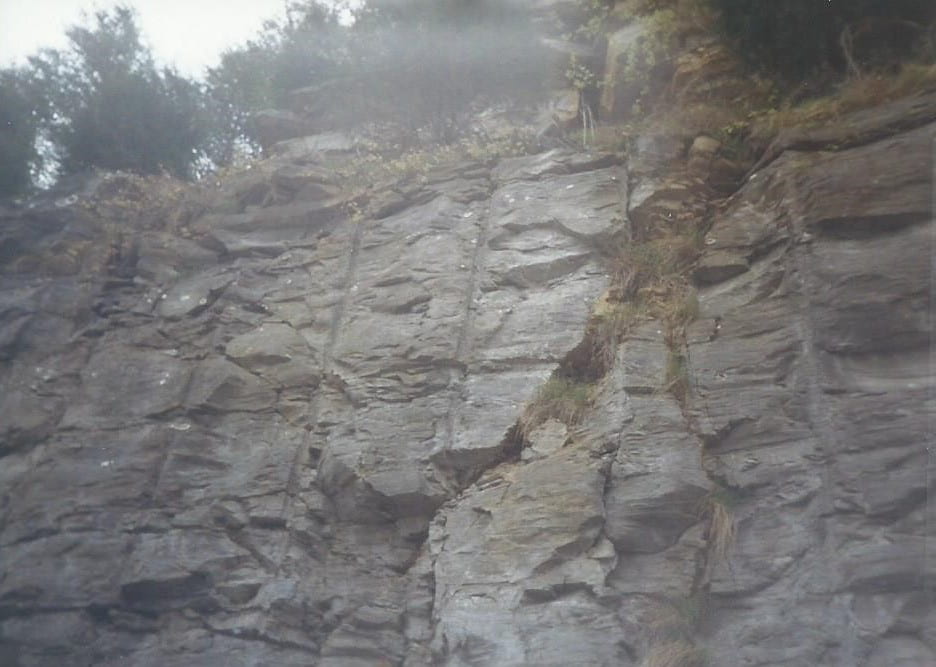
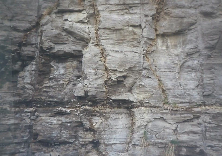
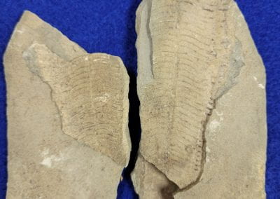
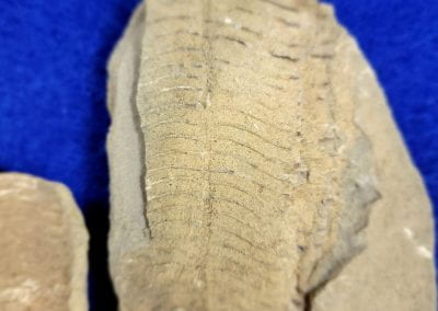
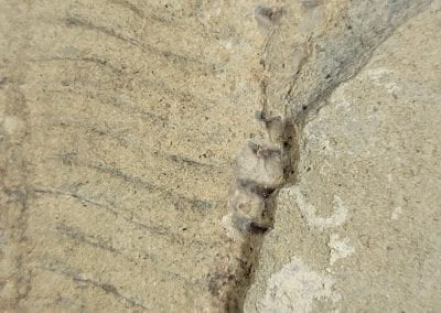
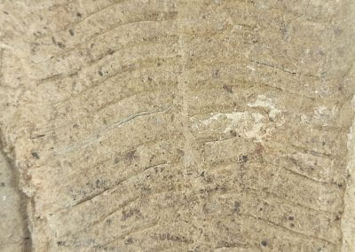
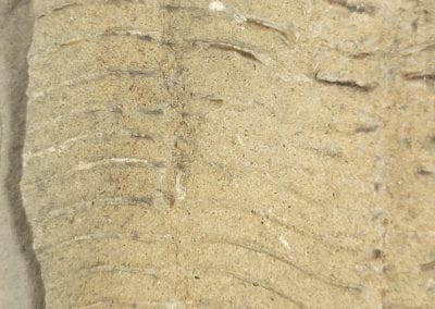
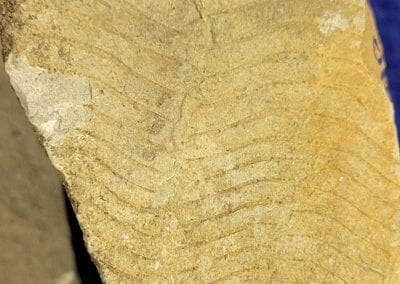
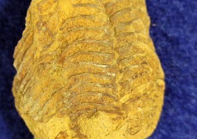
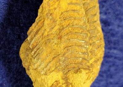
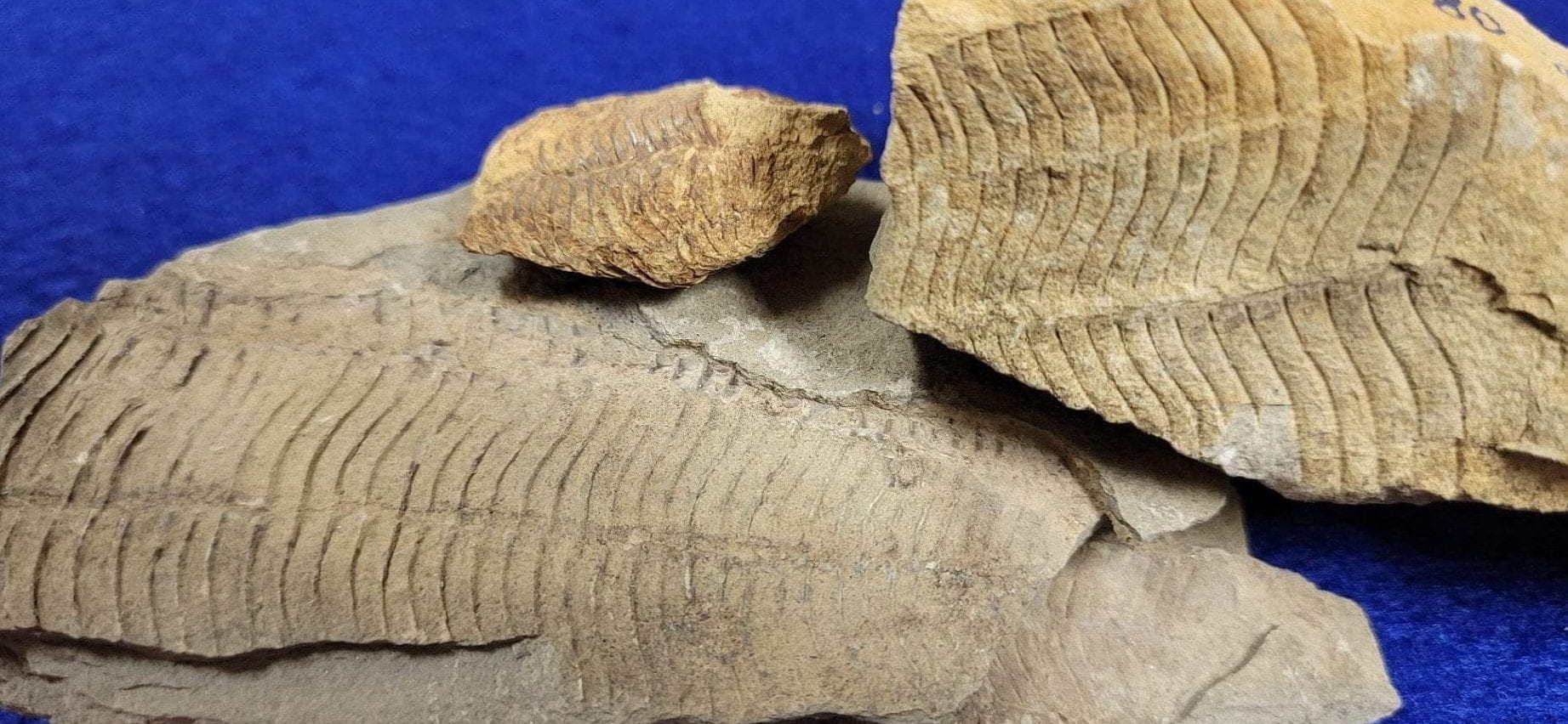
The three conulariid specimens below were collected from a Cumberland Parkway road cut in Kentucky. (1993 L. Curtis)
Stratigraphy of Location
The lithostratigraphy consists of a thick layer of limestone (Middle to Late Mississippian, 346-323.2ma) inclusive of chert nodules, dolostone, and an argillaceous sandstone band (epeiric sediments). Blue River Group/Ste. Genevieve and St. Louis Limestone Formations (Photos L. Curtis)
Conulariid Specimens
(Collected and photographed by L. Curtis, 1993)
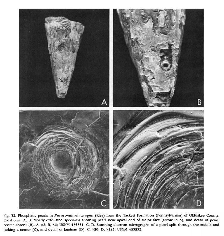
Conulariid Pearls
Babcock, L.E. (1990). Conulariid Pearls. pp. 68-71 IN: Evolutionary Paleobiology of Behavior and Coevolution. Elsevier Scientific Publishing, 725 pp.
“Pearls are reported here for the first time in the extinct animal phylum Conulariida. Pearls (Fig. 52A-D) are rare and have been found in only two species, Paraconularia crustula (White) from the Pennsylvanian of Johnson County, Kansas (USNM 435350), and P. magna (Ries) from the Pennsylvanian of Okfuskee County, Oklahoma (USNM 435351, 435352). They are found adhering either to inner layers of integument in examples having a partially exfoliated exoskeleton (Fig. 52A, B,) or to steinkerns where the exoskeleton has been removed. Examined pearls seem to be of the blister type but some may have initially been cyst pearls that were later incorporated into the inner layers of the exoskeleton.”
“The maximum number of pearls found in one specimen is three and the largest such structure is 2 mm in diameter. Conulariid pearls are composed of numerous fine concentric laminae (Fig. 52C, D) of calcium phosphate. Investigation under high magnification revealed that the laminae are cryptocrystalline. Evidently, these phosphatic concretions formed by the sequential deposition of thin layers of collophane around foreign irritants that affected either the internal exoskeletal surface or soft tissues that bounded it. The irritants that stimulated the formation of pearls are not known. By analogy with pearls formed naturally by present-day mollusks, however, it is probable that many developed around parasites or boring endobionts. Sediment grains may also have served as nuclei. The presence of pearls in conulariids supports evidence (Babcock et al., 1987) that a mantle or mantle-like tissue bounded the internal surface of the exoskeleton and was responsible for its secretion.”
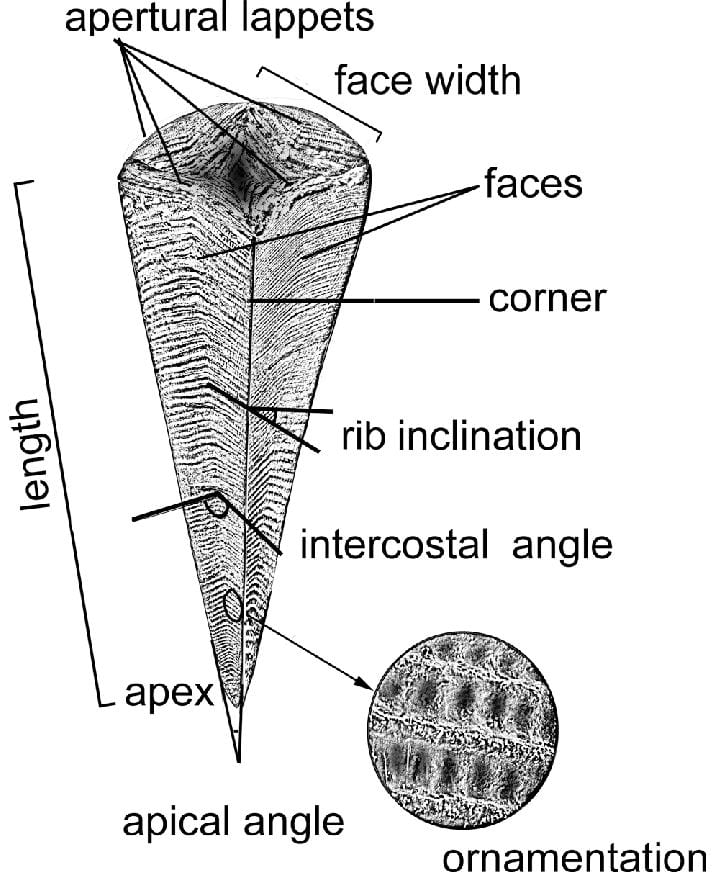
Exoskeletal Structure and Living Representation of Conulariids
Schematic drawing of the conulariid described with main morphological elements (after Sendino, 2006)
Conulariid Thecal Closures and Ornamentation
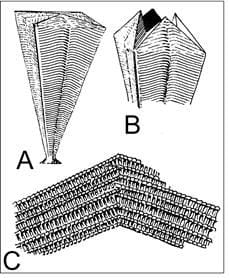
Figure 1 – Lucas, Spencer G. 2012. “The Extinction of the Conulariids” Geosciences 2, no. 1: 1-10. https://doi.org/10.3390/geosciences2010001
Figure 2 – Simoes, Marcello & Rodrigues, Sabrina & Leme, Juliana & H, Van. (2003). Some Middle Paleozoic Conulariids (Cnidaria) as Possible Examples of Taphonomic Artifats. Journal of Taohonomy. 1. 165-186.
Figure 1. (A) Generalized drawing of the conulariid test; (B) Distal end with triangular flaps “lappets” raised; (C) Detail of part of exterior of test showing ornamentation.
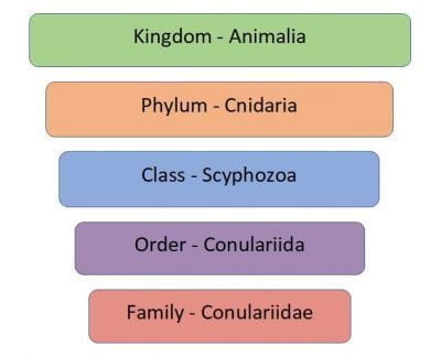
Currently Accepted Taxonomy for Conulariids
Cladistic Analysis of the Suborder Conulariina Miller and Gurley, 1896 (Cnidaria, Scyphozoa; Vendian–Triassic) by Juliana de Moraes Leme, Marcello Guimaraes Simoes, Antonio Carlos Marques and Heyo van Iten – June 2007
Successive Weighting Tree for Conulariids
Cladistic Analysis of the Suborder Conulariina Miller and Gurley, 1896 (Cnidaria, Scyphozoa; Vendian–Triassic) by Juliana de Moraes Leme, Marcello Guimaraes Simoes, Antonio Carlos Marques and Heyo van Iten – June 2007
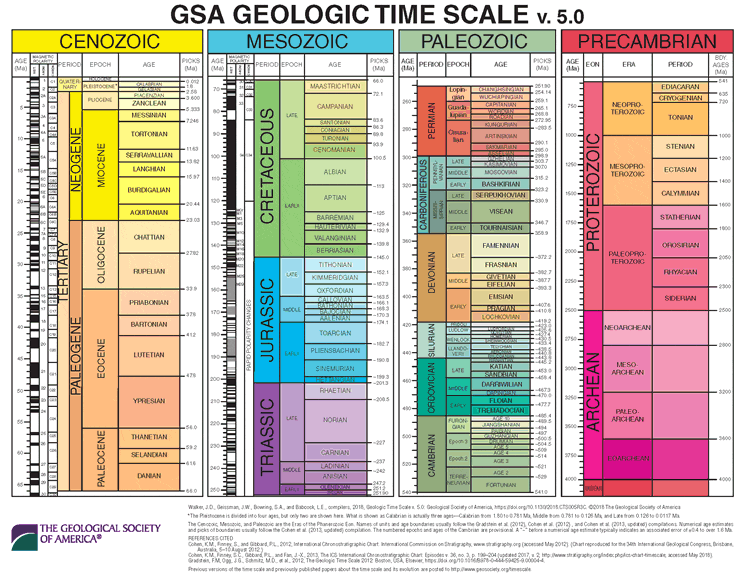
Geological Society of America Geologic Time Scale (2018)(v. 5.0)
Click the Geologic Time Scale for a pdf and better view.

